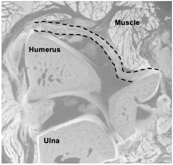
Images below are from our main lab projects:
Quantitative Polarization Imaging (QPLI)
Post-traumatic Joint Contracture (PTJC)
Multiscale Tendon Mechanics (Tendon)
Abdominal Hernia (Hernia)
Hernia
Tendon
Tendon
QPLI
Tendon
Tendon
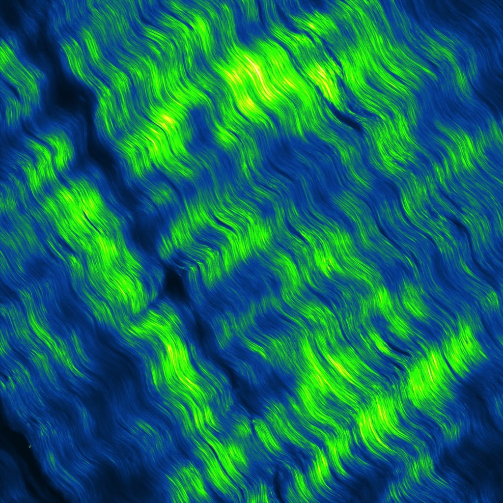
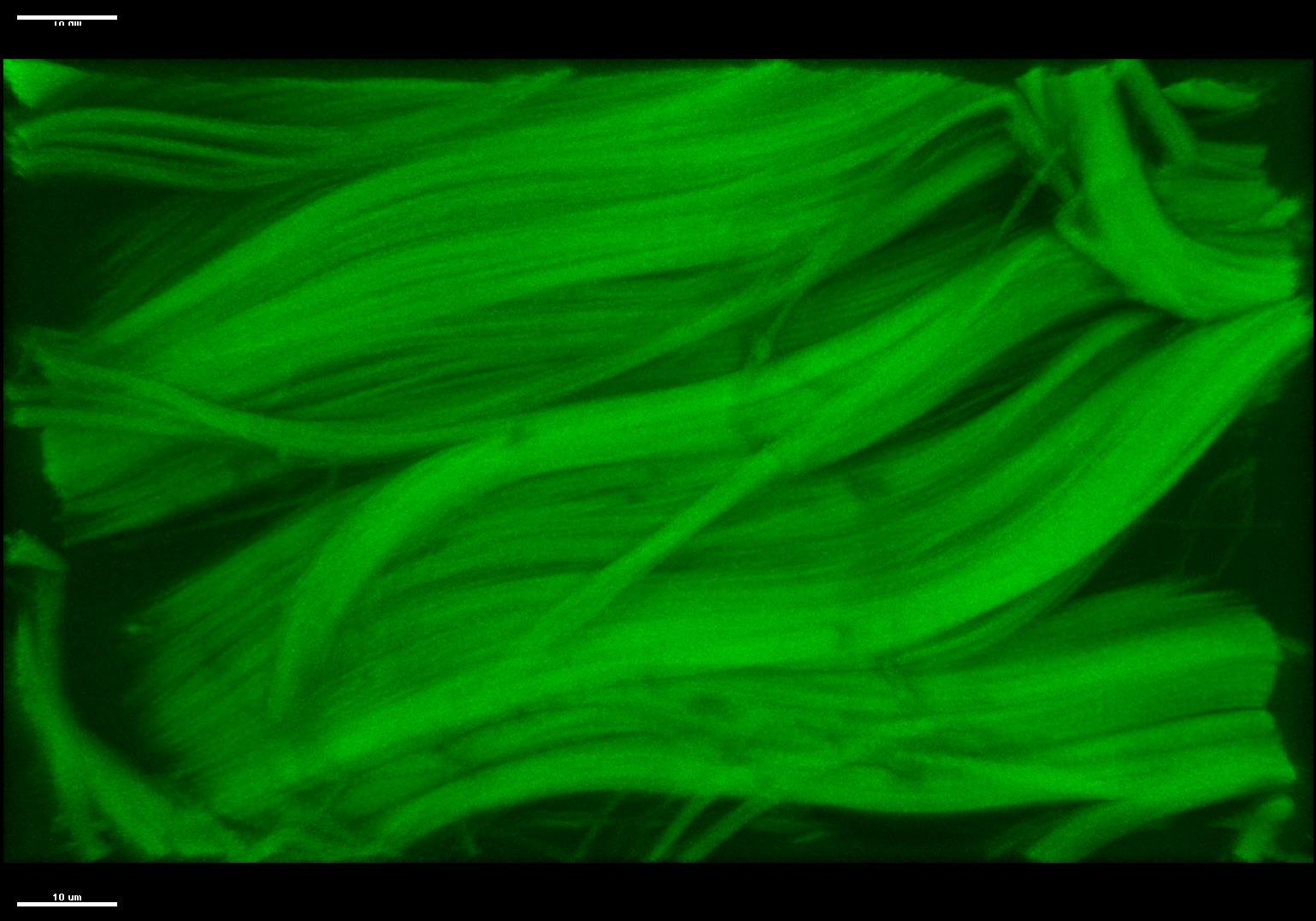
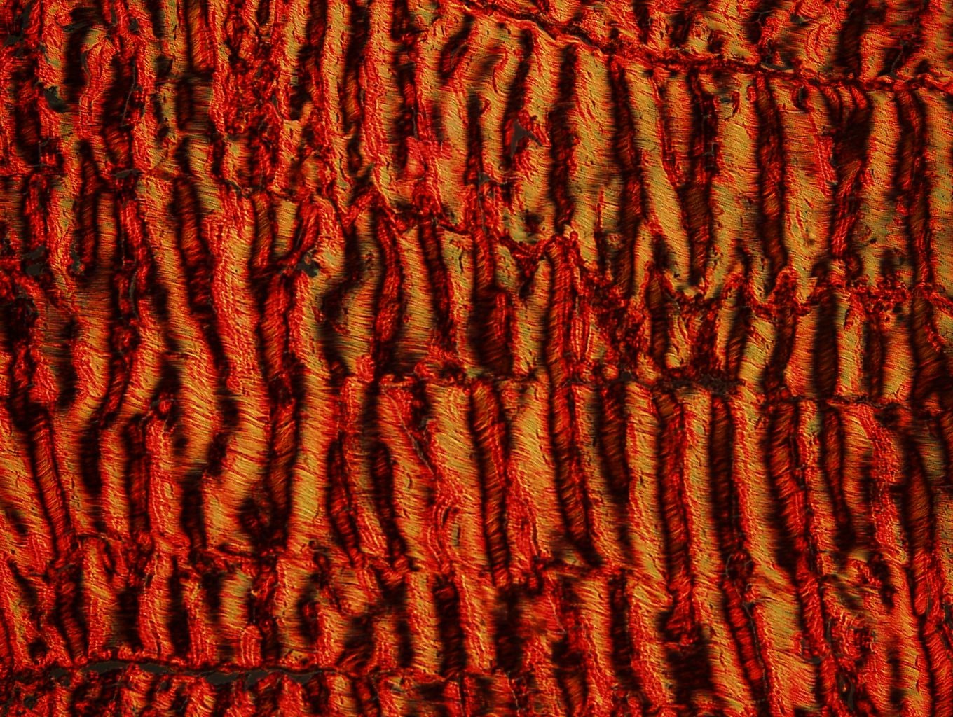
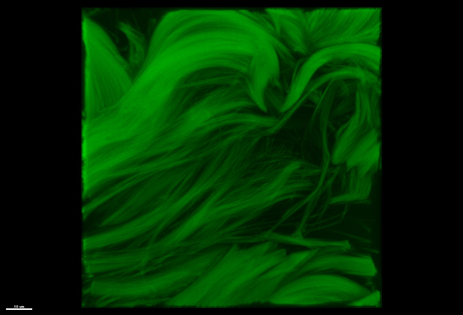
Pseudo colored second harmonic generation images of bovine flexor tendon.
PTJC
QPLI
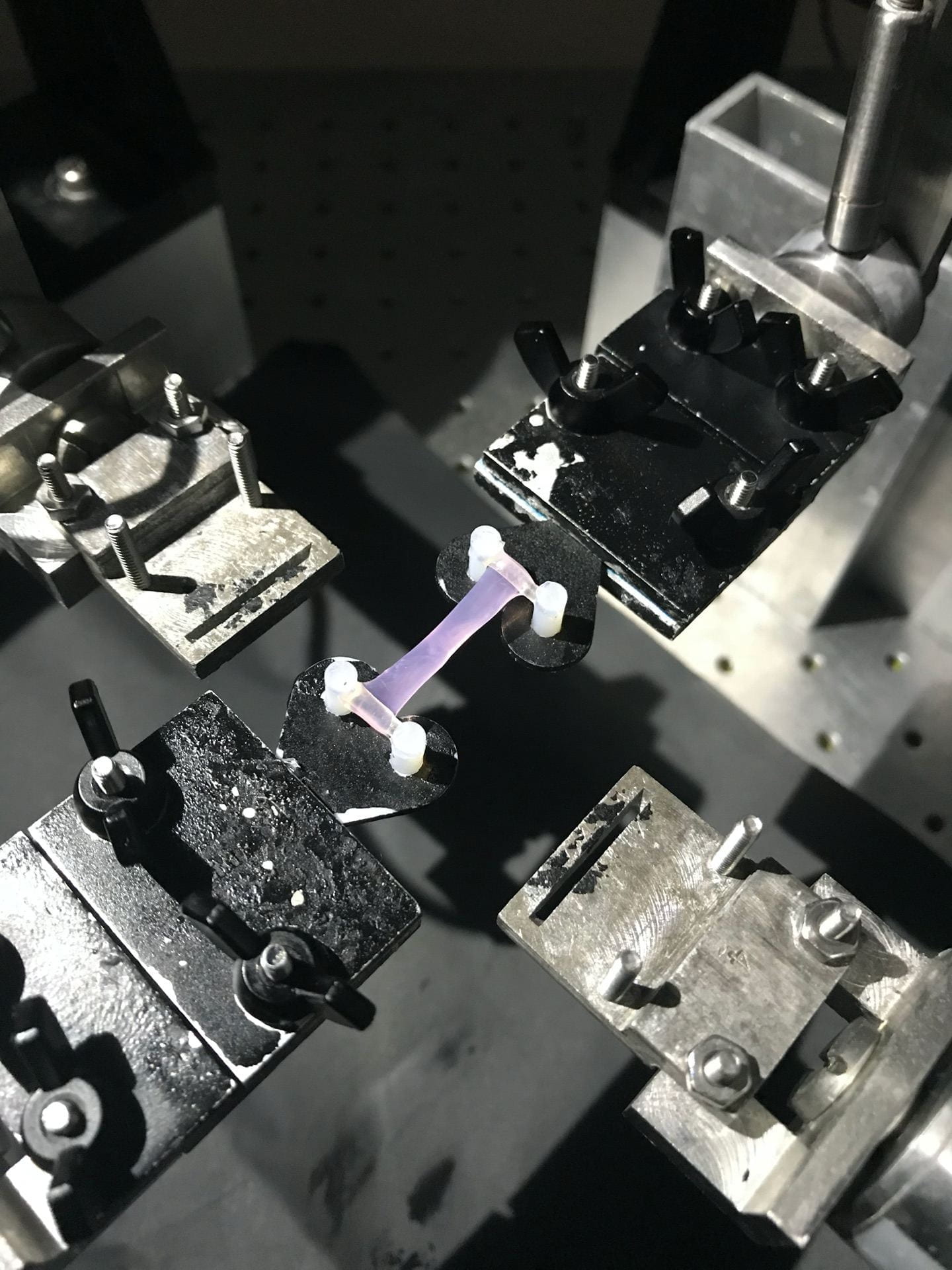
Fibroblast-seeded collagen gel tissue analog with highly aligned microstructure loaded in biaxial testing apparatus and ready for rQPLI.
Tendon
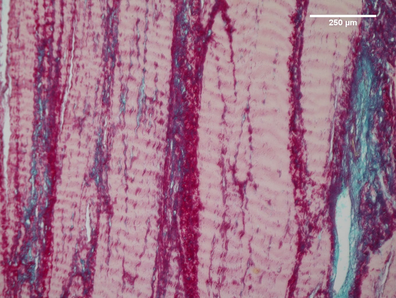
Alcian blue and pico serious red histological staining of bovine deep digital flexor tendon used to visualize proteoglycan content (blue) and collagen content (pink).
Tendon
PTJC
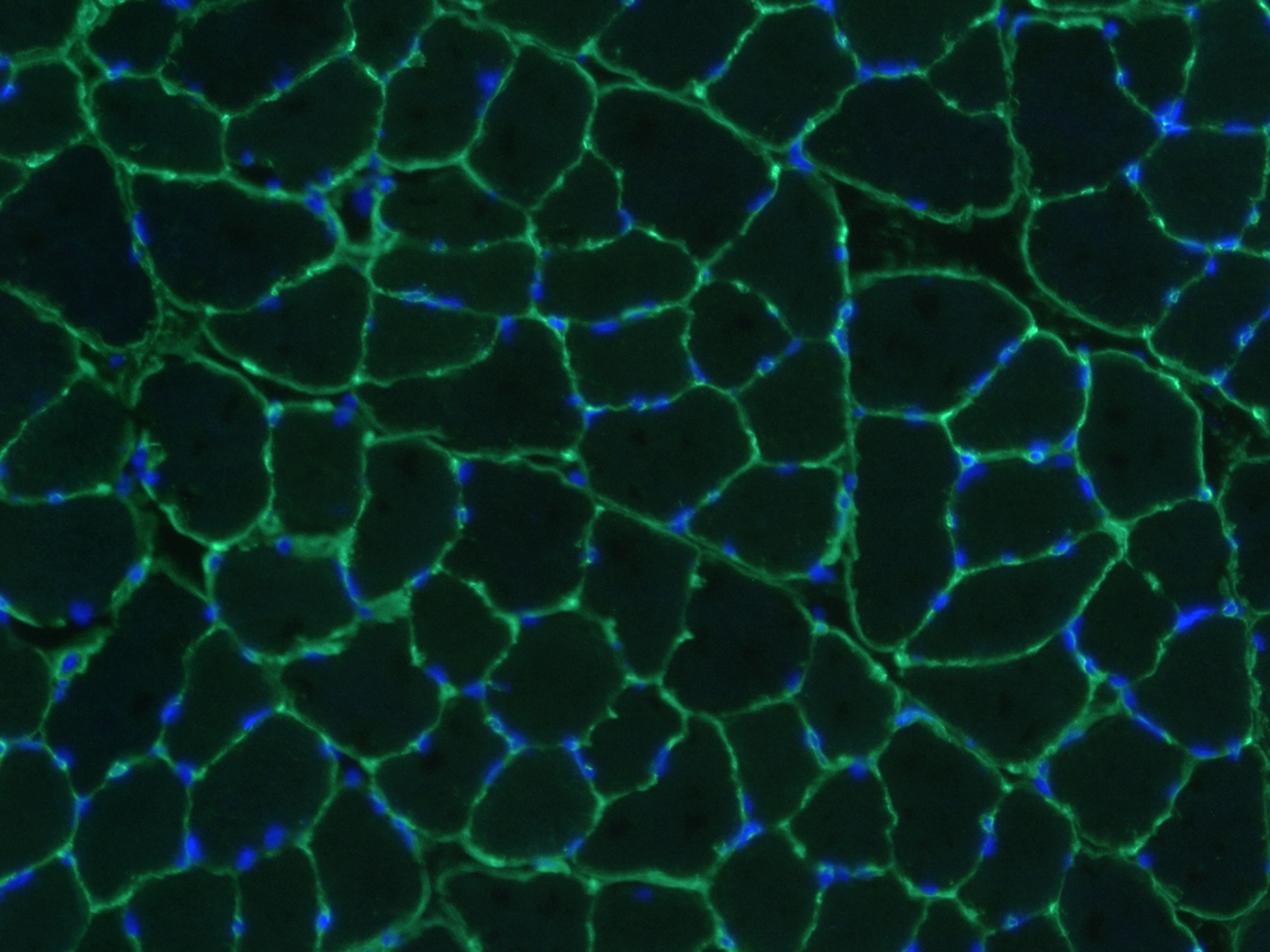
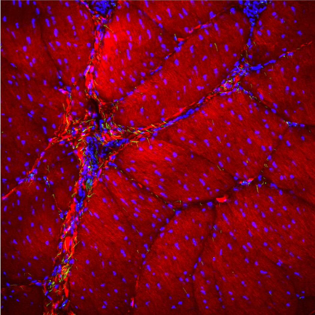
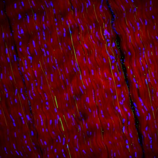
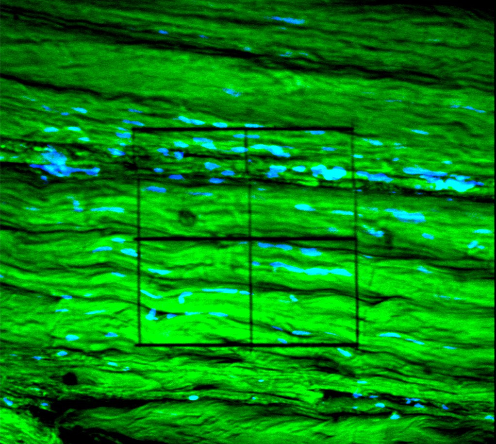
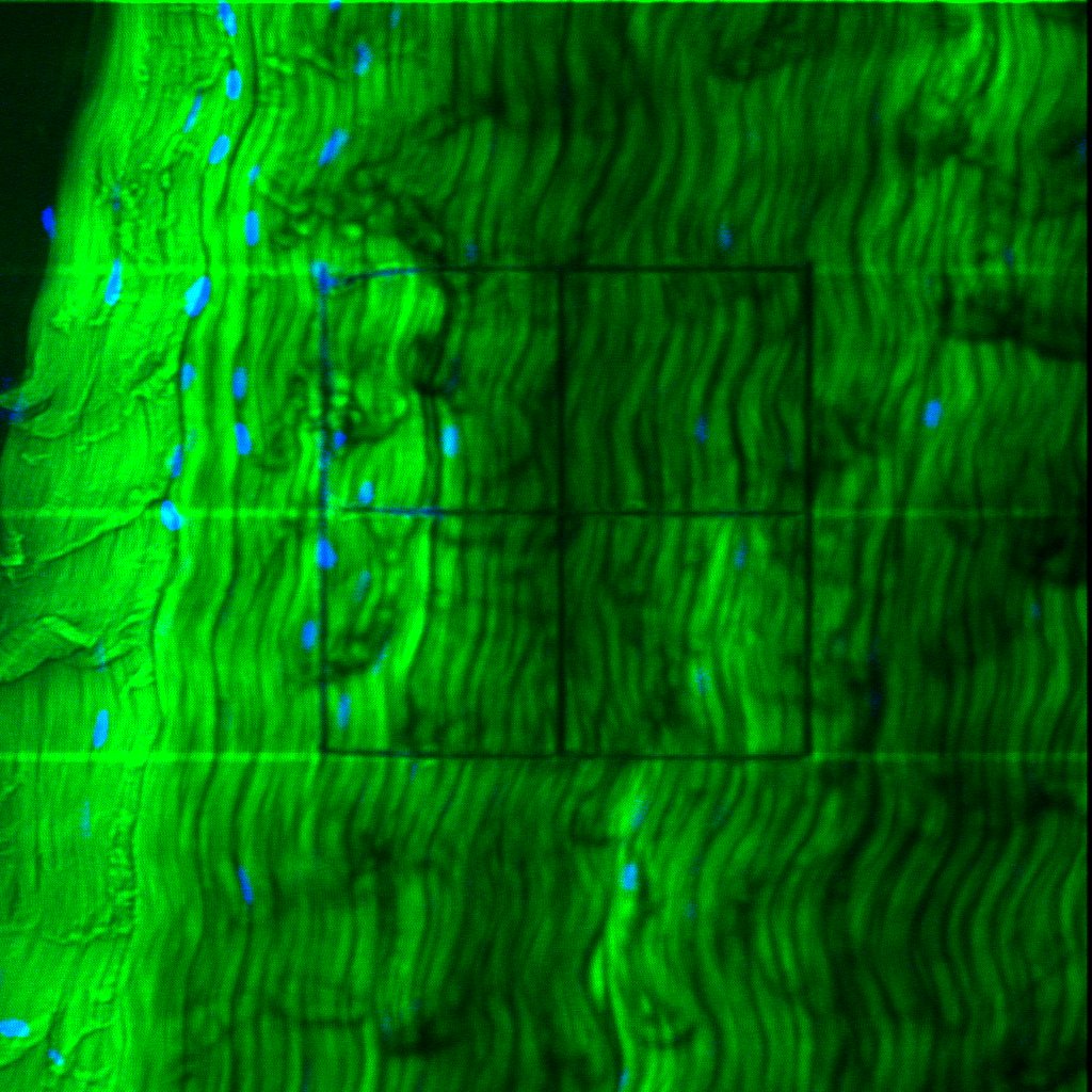
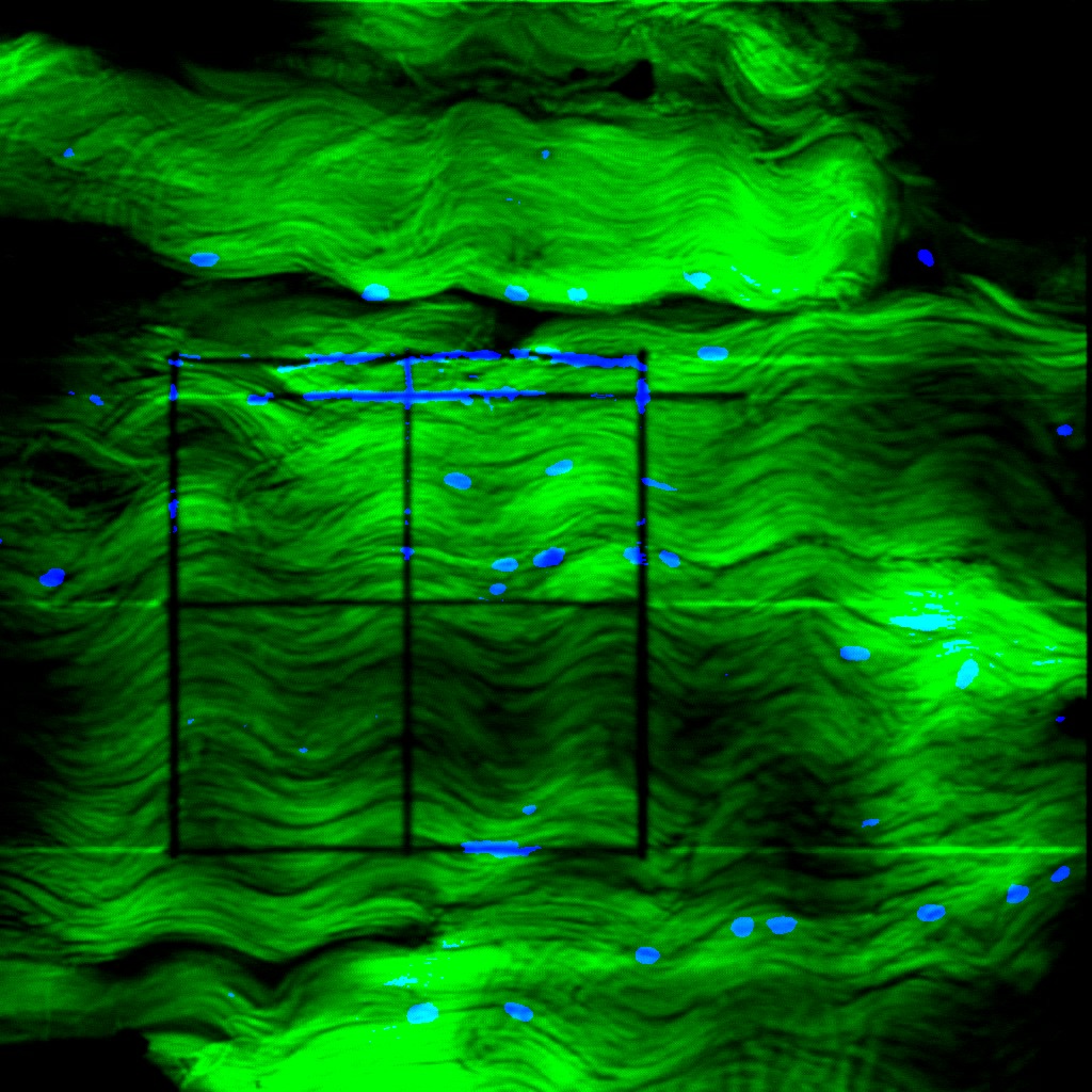
Muscle section immunolabeled with laminin (green) and DAPI (blue).
Tendon
Tendon
PTJC
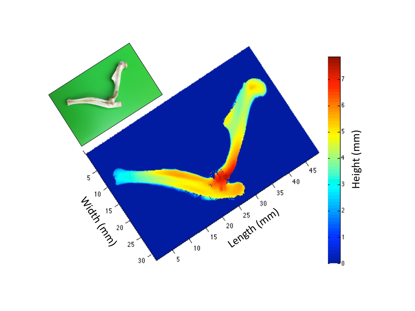
Bones of an elbow scanned using a non contact laser scanner.
PTJC

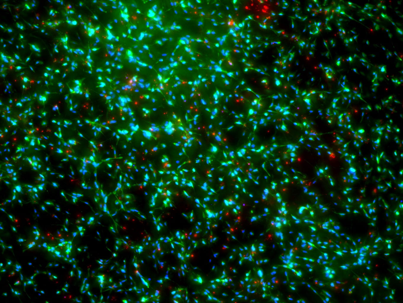
Rat primary adipose stem cell soft primed on 1 kPa hydrogel for two weeks then transferred to 120 kPa hydrogel for one day and immunolabeled with phalloidin (green), YAP (red), and DAPI (blue).
Tendon
PTJC
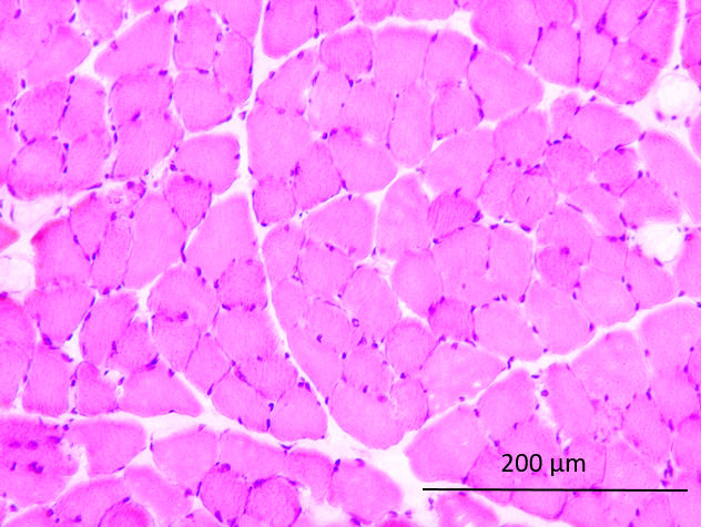
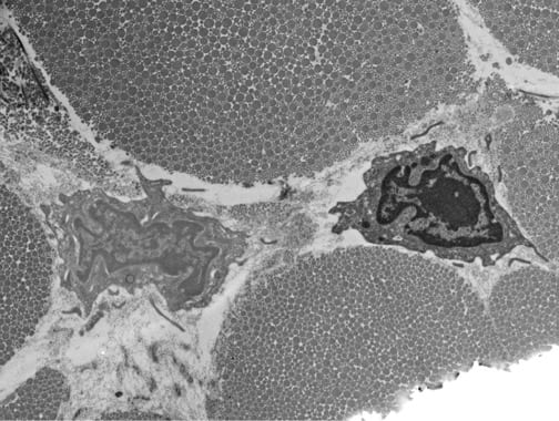
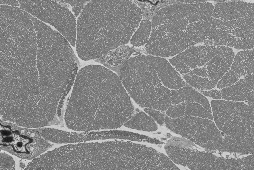
Muscle section stained with hematoxylin and eosin.
Tendon
Tendon
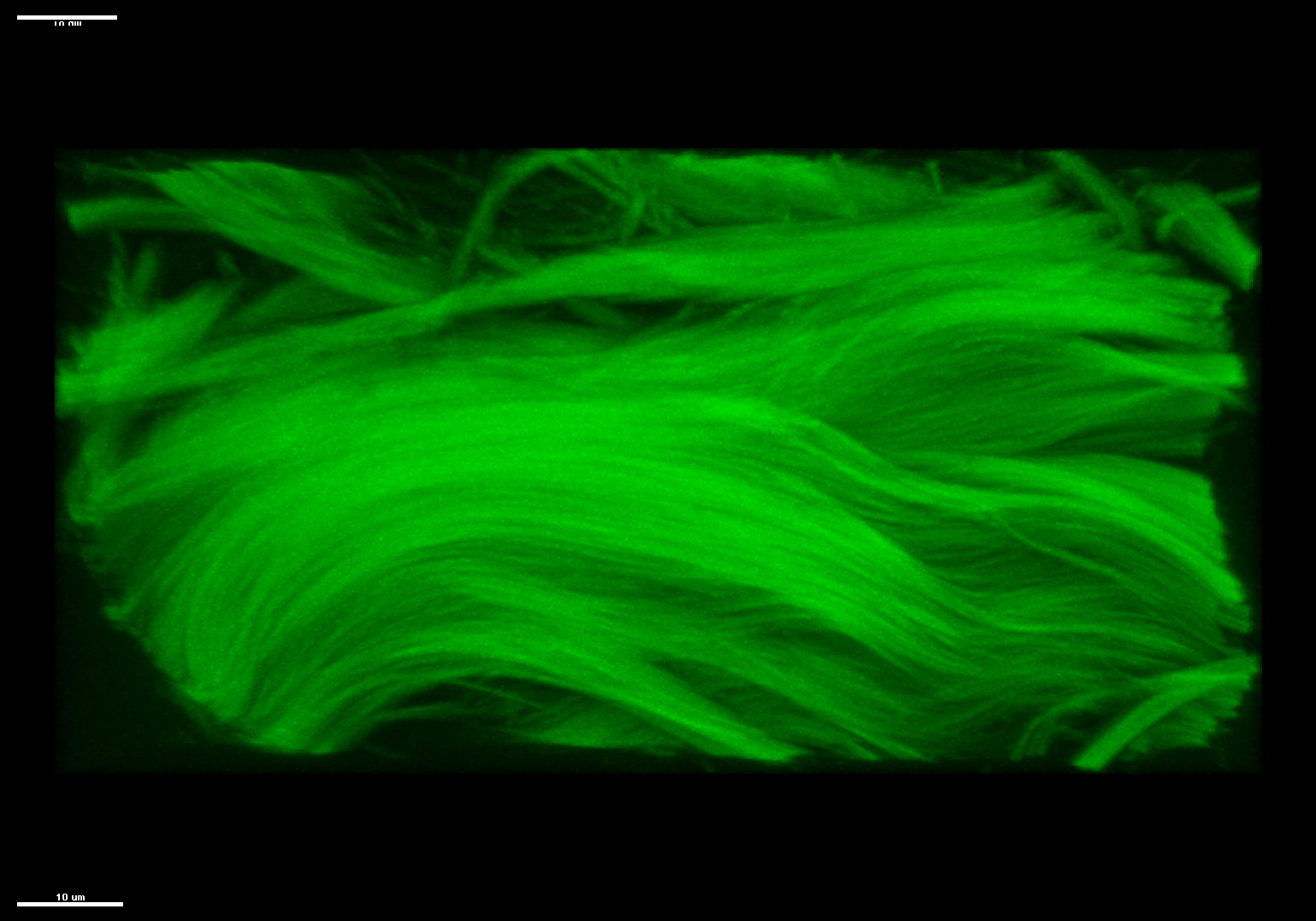
PTJC
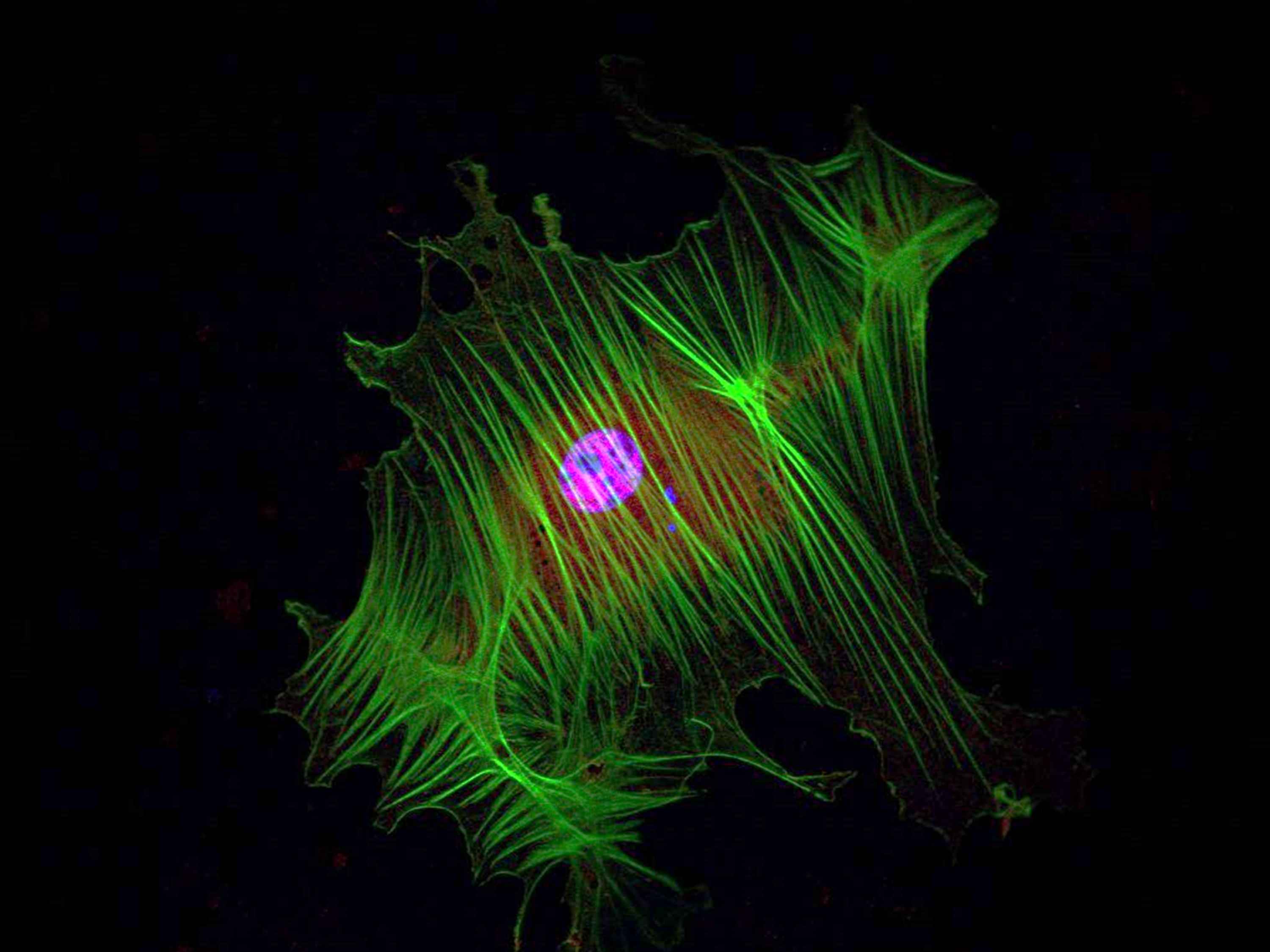
Rat primary adipose stem cell cultured on 120 kPa hydrogel for two weeks and immunolabeled with phalloidin (green), YAP (red), and DAPI (blue).
QPLI
Tendon
QPLI
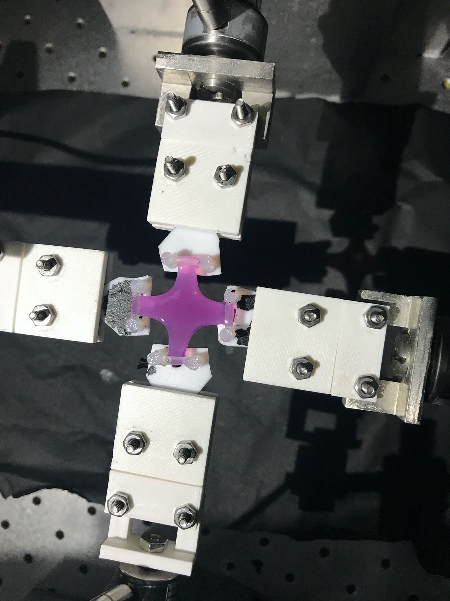
Fibroblast-seeded collagen gel tissue analog with disorganized microstructure loaded in biaxial testing apparatus for rQPLI.
Tendon
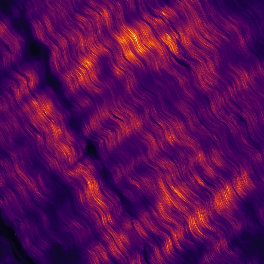
Pseudo colored second harmonic generation images of bovine flexor tendon.
PTJC
QPLI
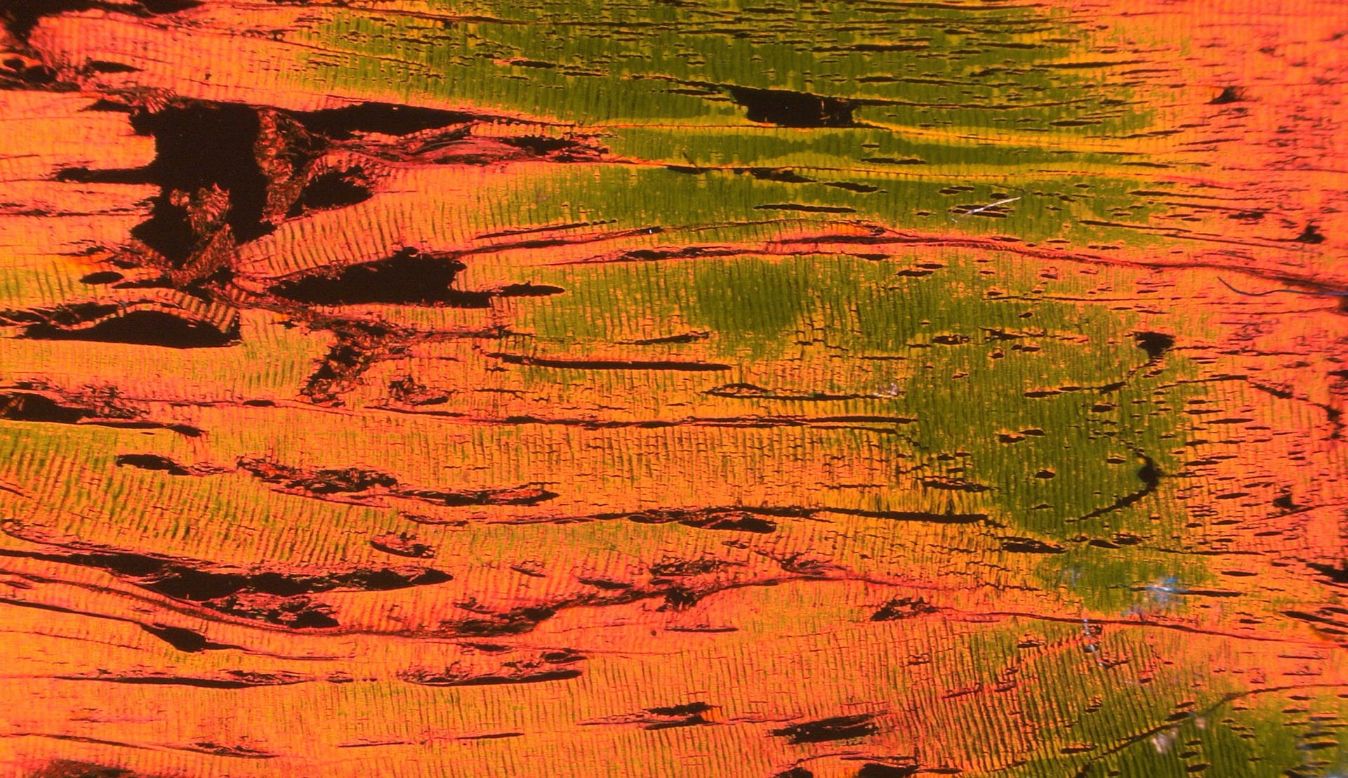
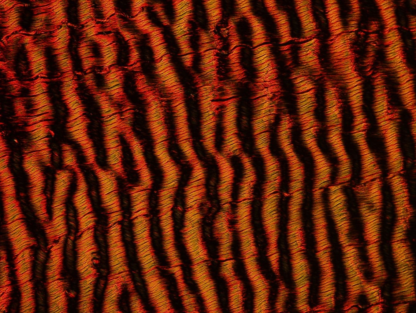
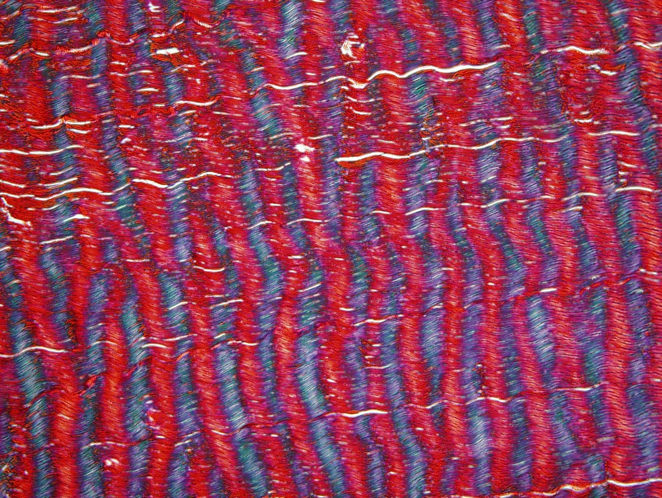
Picrosirius red-stained bovine flexor tendon imaged under polarized light microscopy.
Tendon
PTJC
QPLI
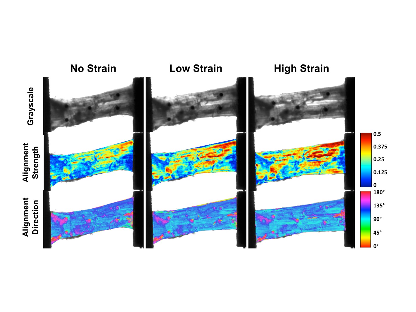
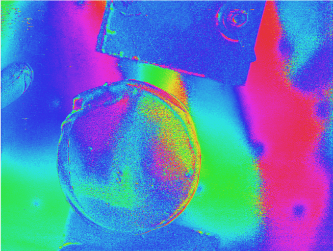
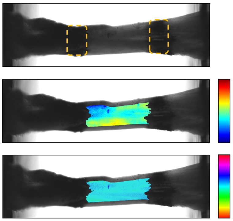
Example of what is displayed when imaging a sample with the polarization camera.
Tendon
QPLI
Tendon
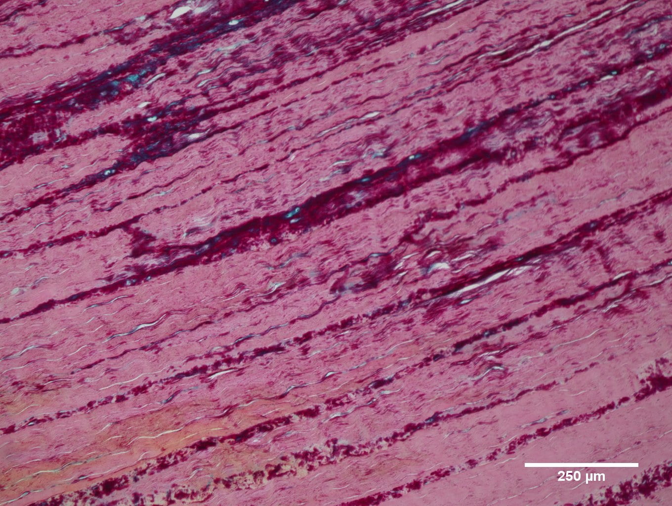
Alcian blue and pico serious red histological staining of bovine deep digital flexor tendon used to visualize proteoglycan content (blue) and collagen content (pink).
PTJC
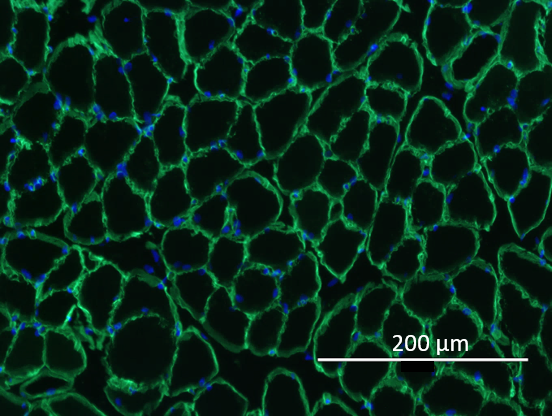
Muscle Section immunolabeled with laminin (green) and DAPI (blue).
Tendon
Tendon
Tendon
Tendon
Tendon
PTJC
Joint section pseudo-colored to create a fun image!

