Images below are from our main lab projects:
Quantitative Polarization Imaging (QPLI)
Post-traumatic Joint Contracture (PTJC)
Multiscale Tendon Mechanics (Tendon)
Abdominal Hernia (Hernia)
Hernia

Surface meshes and contour maps are created to visualize the geometry and deformation of the abdominal wall at the normal intra-abdominal pressure (left) and the maximum intra-abdominal pressure (right) of the applied cough.
Tendon
Transverse section of bovine long digital extensor tendon showing collagen (red), elastin (green), and cell nuclei (blue) highlighting the elastin-rich extracellular matrix.
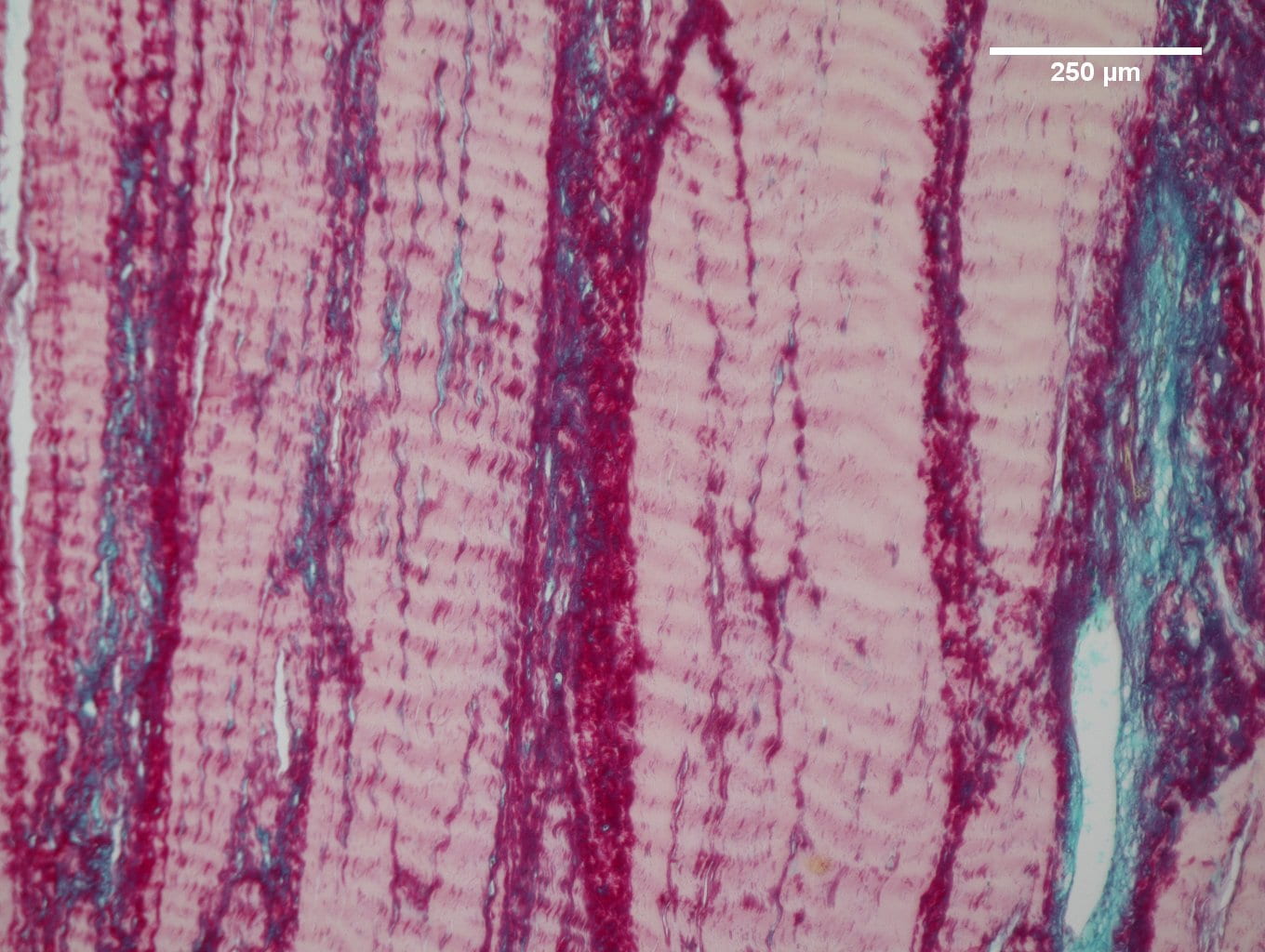
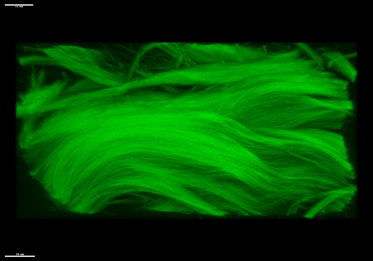
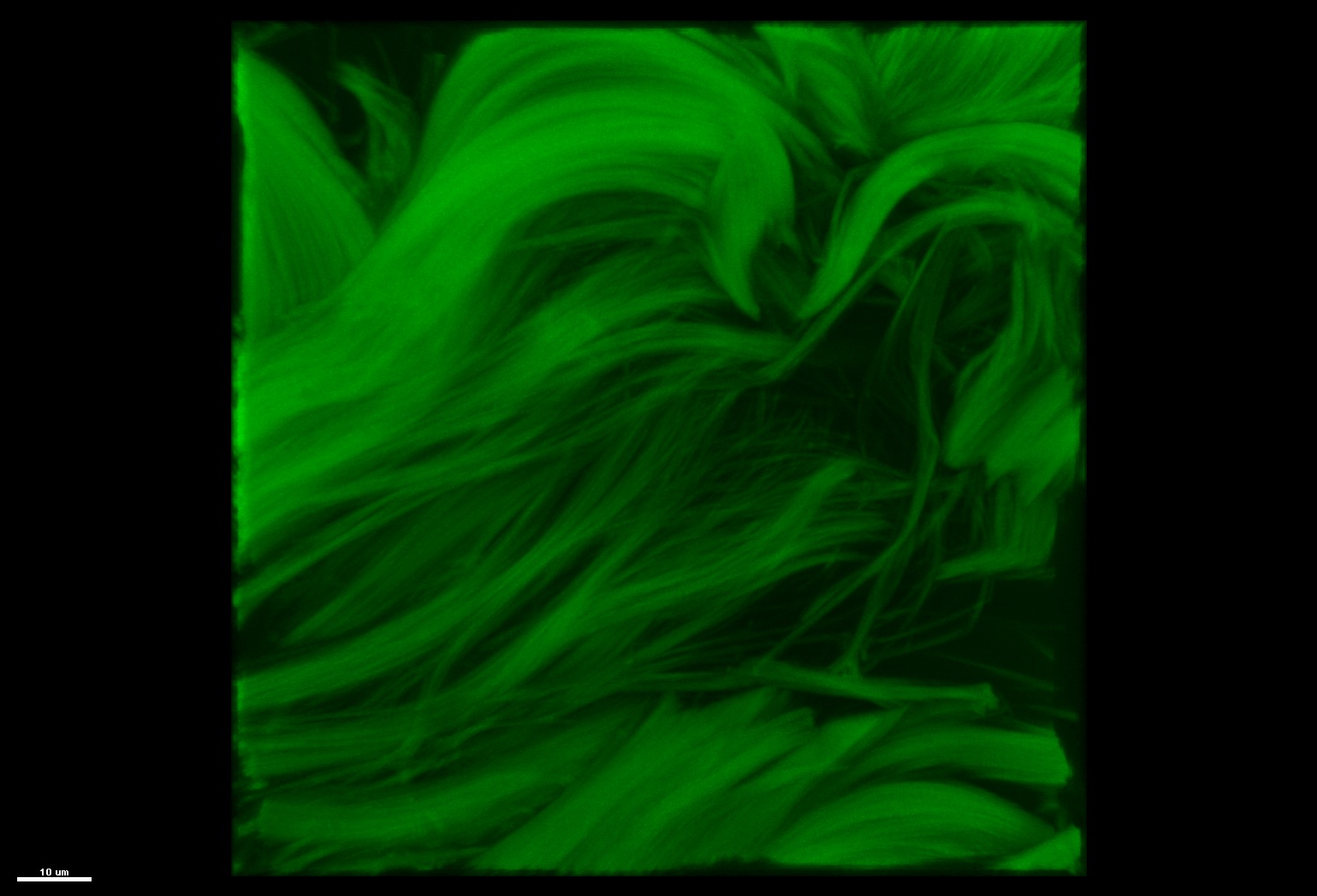
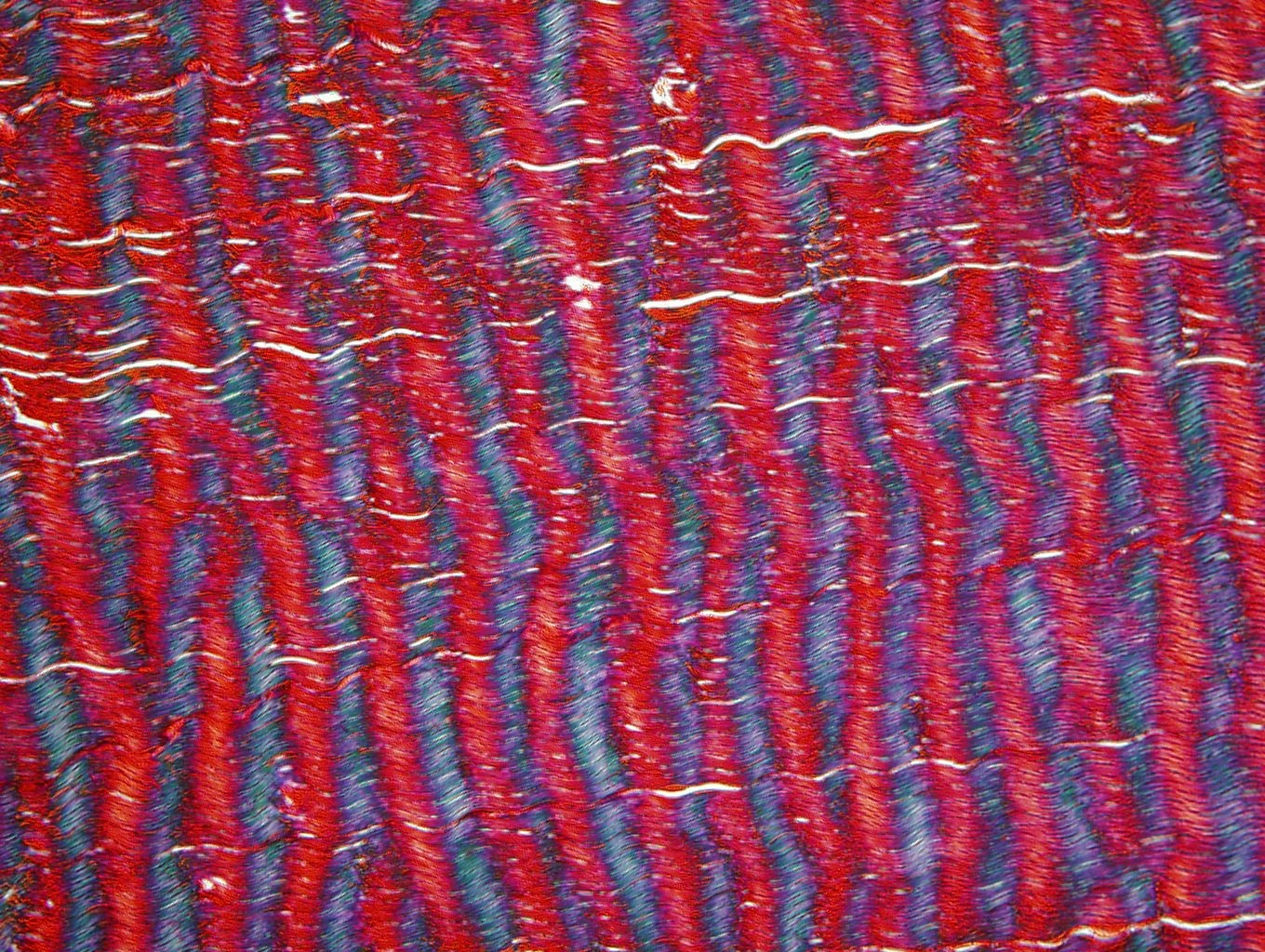
Tendon
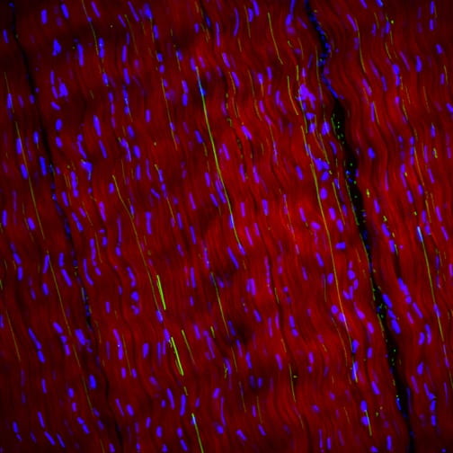
Longitudinal section of bovine superficial digital flexor tendon showing collagen (red), elastin (green), and cell nuclei (blue) highlighting the aligned collagen and elastic fibers.
QPLI

“Polarized Light Art” created using plastic, tape, and a polarized light source (computer monitor) as part of outreach project.
Hernia

Click image above for video of cough simulation on our hernia box.
Tendon
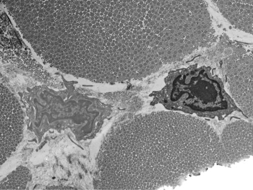
Longitudinally sectioned transmission electron micrograph of murine tibialis anterior tendon highlighting the complex pericellular matrix.
PTJC

Paw print image from gait analysis.
QPLI
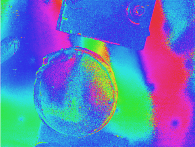
Circular collagen gel placed on a plastic Petri dish and imaged using QPLI.
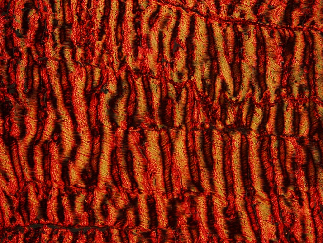
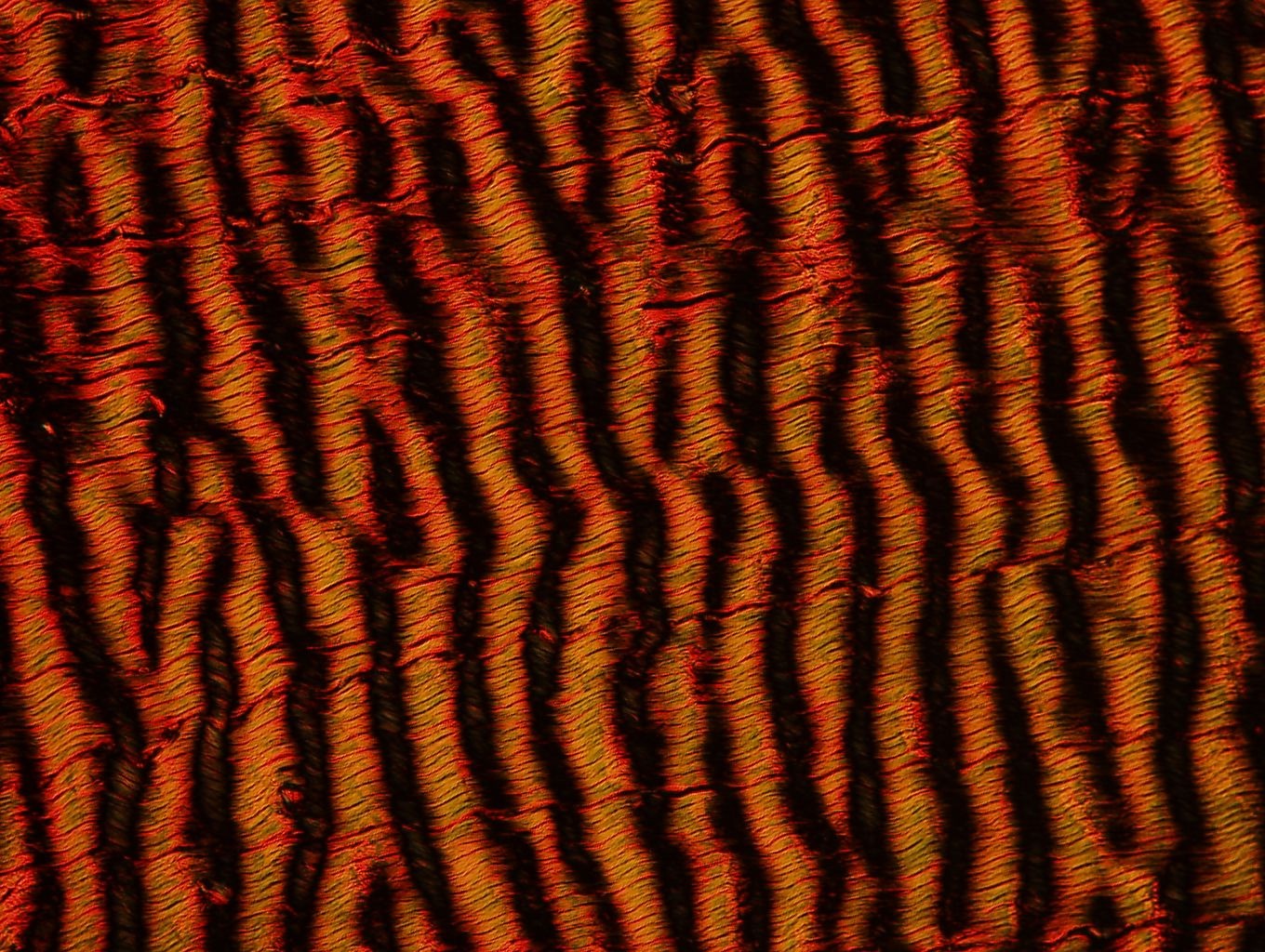
Tendon
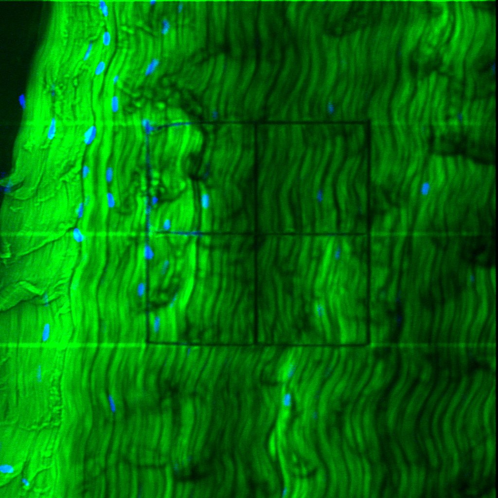
Pseudo colored second harmonic generation images of bovine flexor tendon.
PTJC
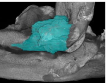
Injured limb lateral collateral ligament (LCL) from contrast enhanced micro-computed tomography imaging technique. Post-processing pseudo-colored the LCL aqua.
QPLI
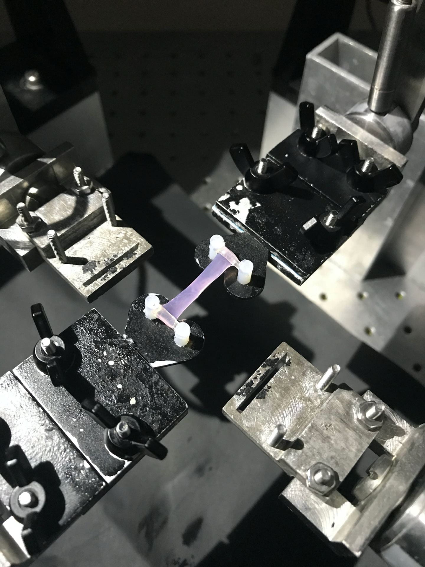
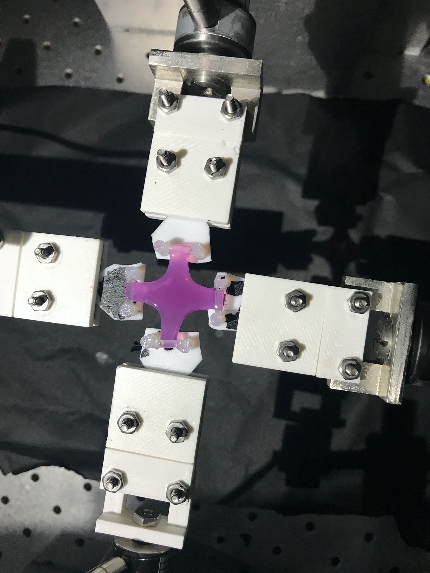
Fibroblast-seeded collagen gel tissue analog with highly aligned microstructure loaded in biaxial testing apparatus and ready for rQPLI.
Tendon
Alcian blue and pico serious red histological staining of bovine deep digital flexor tendon used to visualize proteoglycan content (blue) and collagen content (pink).
Tendon
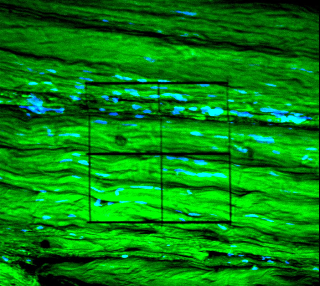
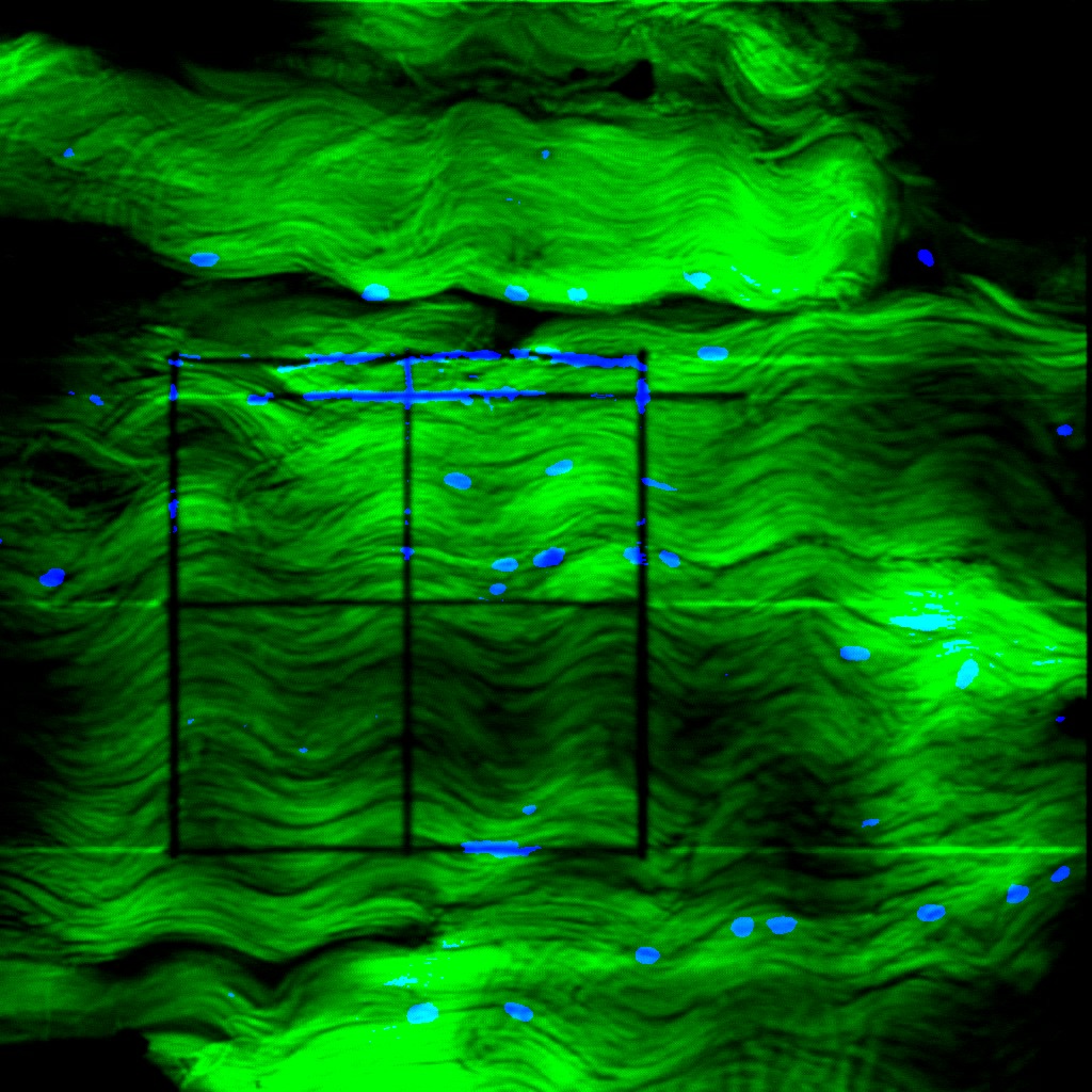
PTJC
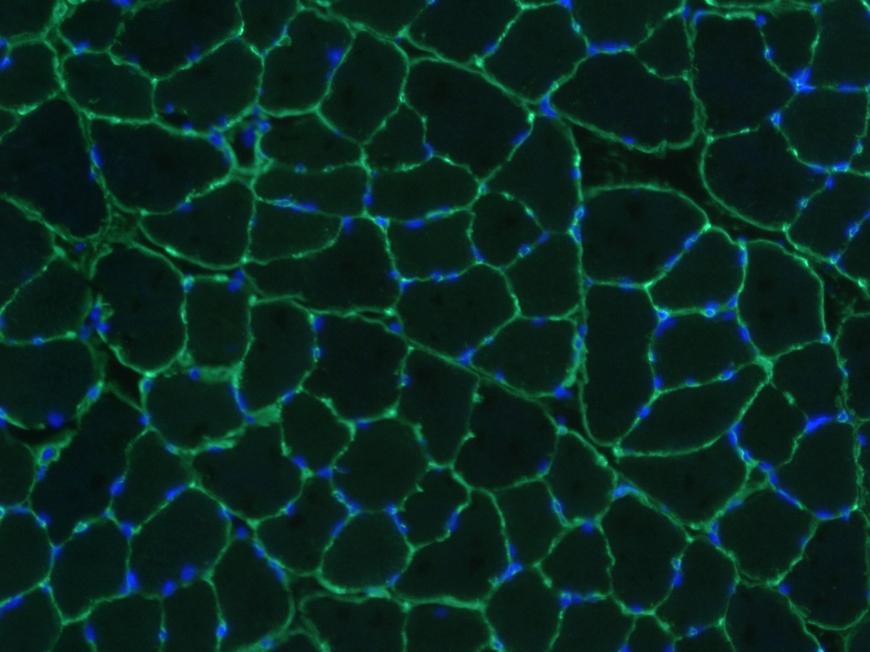
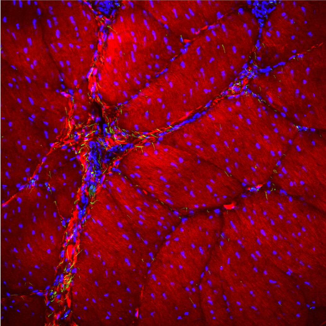
Muscle section immunolabeled with laminin (green) and DAPI (blue).
Tendon
Tendon

PTJC
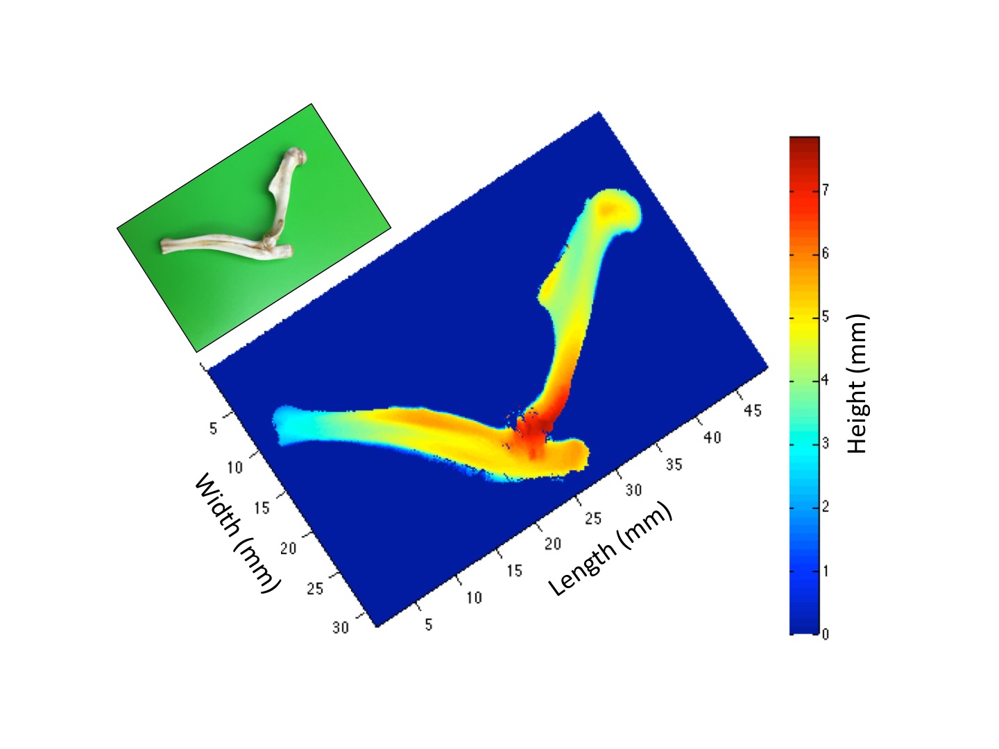
Bones of an elbow scanned using a non contact laser scanner.
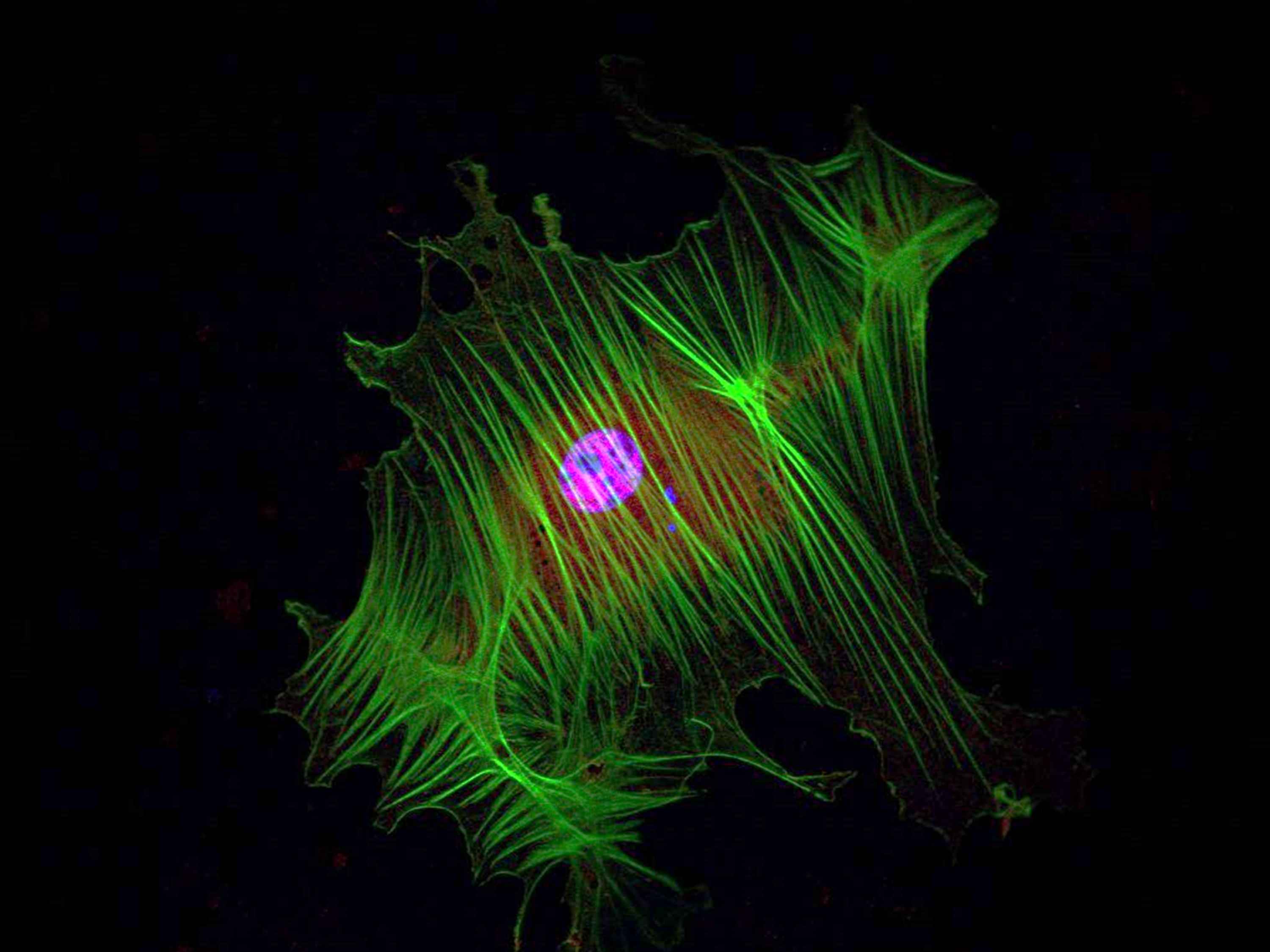
PTJC

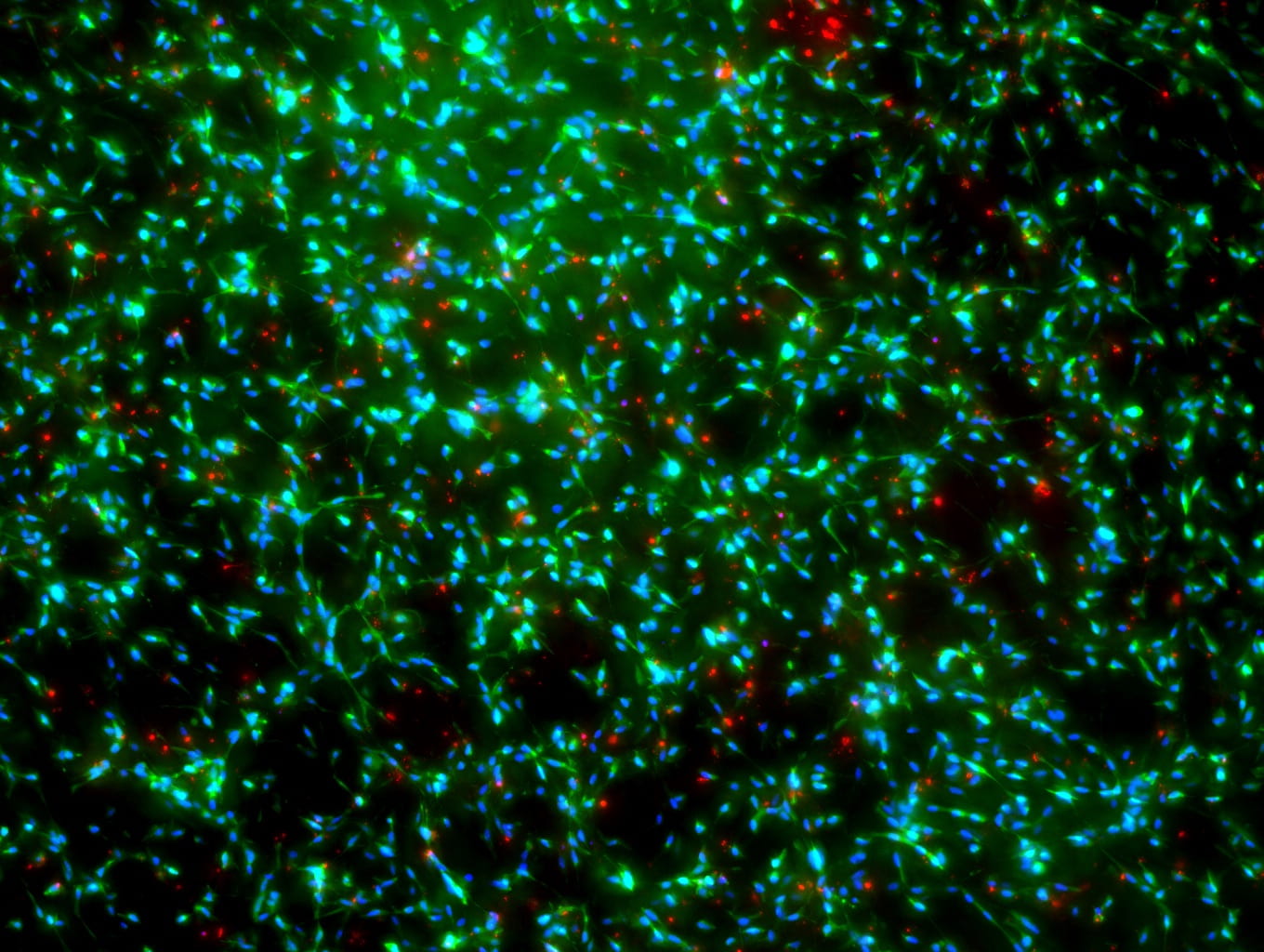
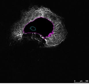
Rat primary adipose stem cell soft primed on 1 kPa hydrogel for two weeks then transferred to 120 kPa hydrogel for one day and immunolabeled with phalloidin (green), YAP (red), and DAPI (blue).
Tendon
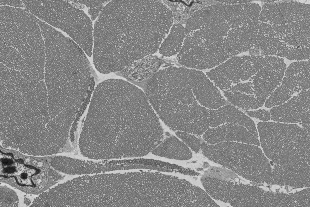
PTJC
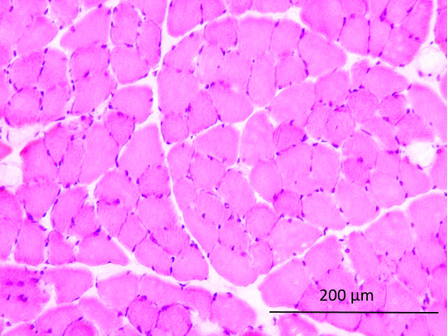
Muscle section stained with hematoxylin and eosin.
Tendon
![]()
Tendon
PTJC
Rat primary adipose stem cell cultured on 120 kPa hydrogel for two weeks and immunolabeled with phalloidin (green), YAP (red), and DAPI (blue).
QPLI

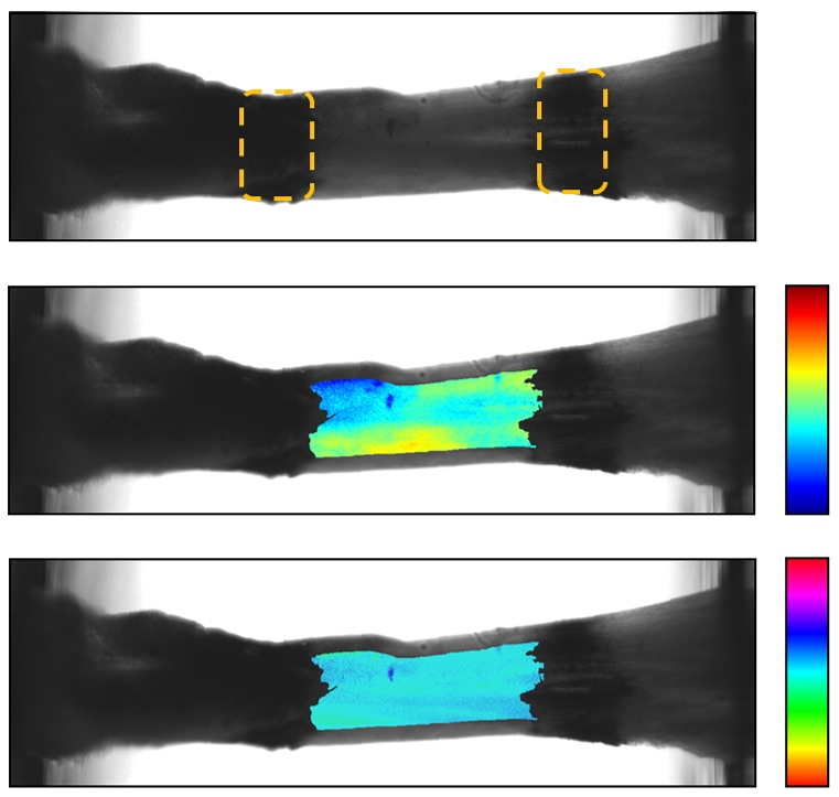
Color maps of polarization metrics from disorganized tissue analogs imaged using QPLI. Left = DoLP. Right = AoP.
Hernia

This device is used to apply a representative coughing force to a porcine abdominal wall.
PTJC
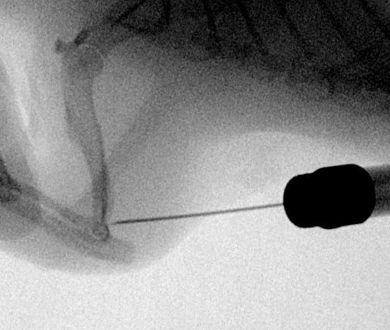
Fluoroscope image of intra-articular rat elbow injections.
Tendon
Transversely sectioned transmission electron micrograph of murine Achilles tendon highlighting the aligned collagen fibrils and elastic fiber.
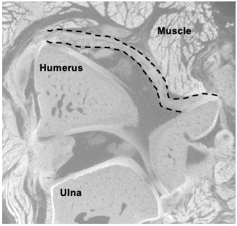
PTJC

Micro-CT of heterotypic ossification around the elbow.
QPLI
Fibroblast-seeded collagen gel tissue analog with disorganized microstructure loaded in biaxial testing apparatus for rQPLI.
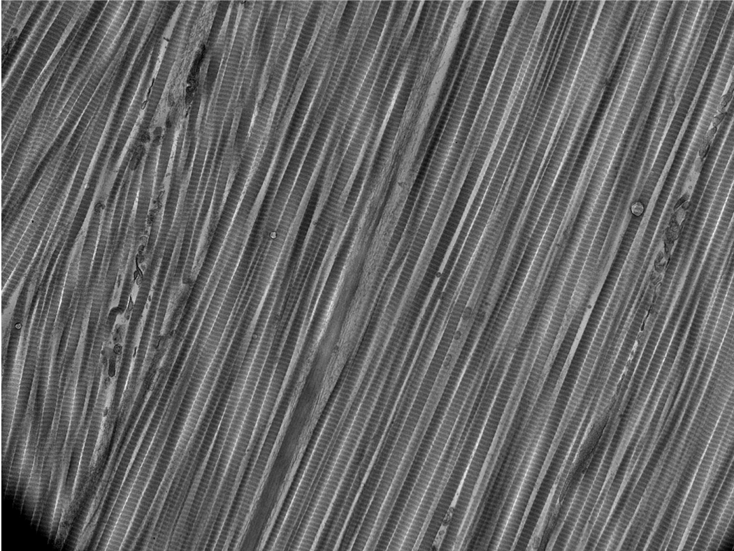
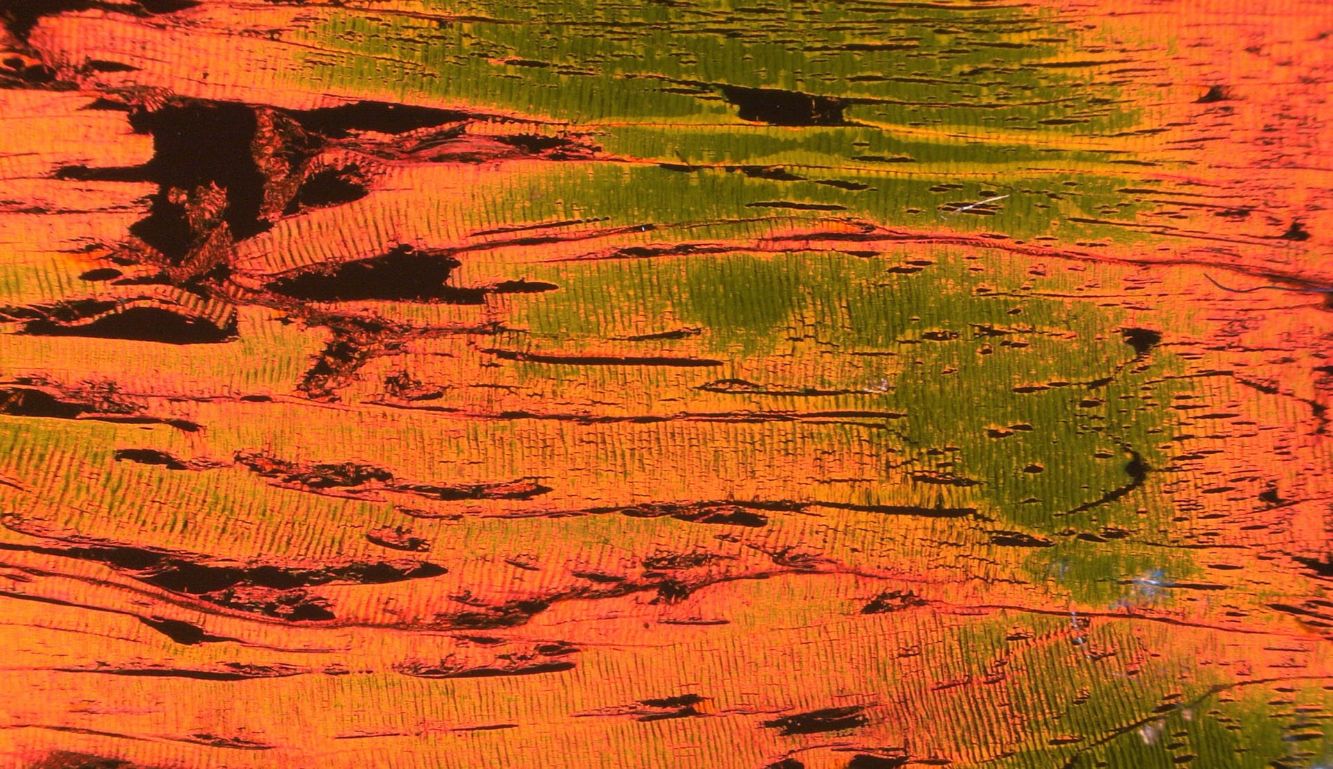
Tendon

Pseudo colored second harmonic generation images of bovine flexor tendon.
PTJC
Rat primary adipose stem cell cultured on 120 kPa hydrogel for two weeks and immunolabeled using FUNCAT extracellular matrix imaging technique.
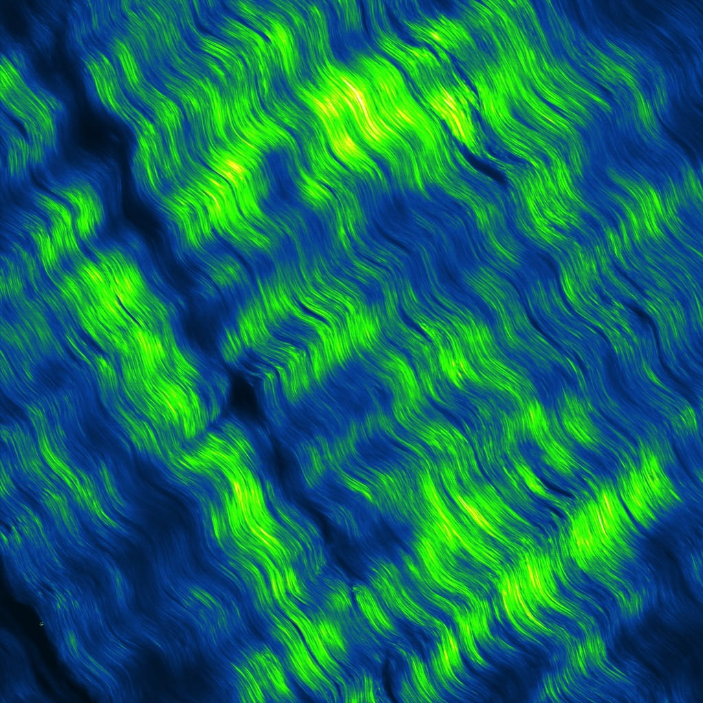
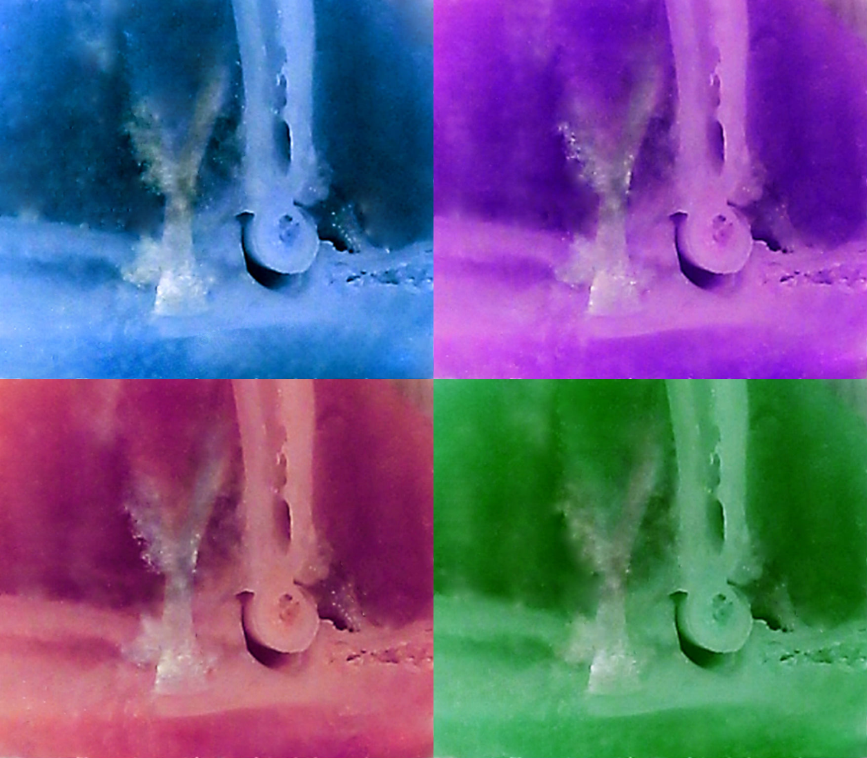
QPLI
Picrosirius red-stained bovine flexor tendon imaged under polarized light microscopy.
Tendon
PTJC
Injured limb anterior capsule from contrast enhanced micro-computed tomography imaging technique.
QPLI
Example of what is displayed when imaging a sample with the polarization camera.
Tendon
QPLI
Tendon
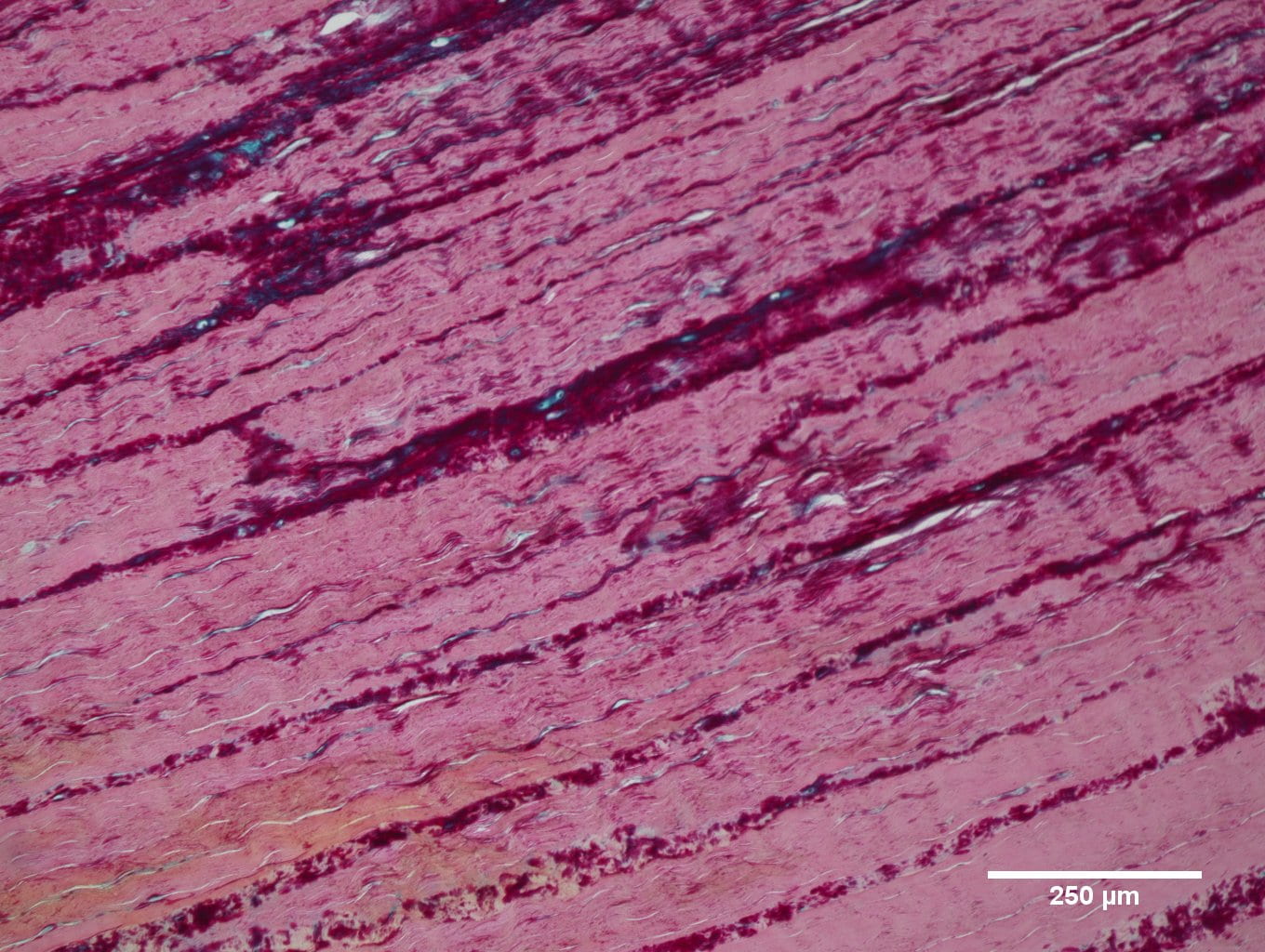
Alcian blue and pico serious red histological staining of bovine deep digital flexor tendon used to visualize proteoglycan content (blue) and collagen content (pink).
PTJC
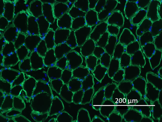
Muscle Section immunolabeled with laminin (green) and DAPI (blue).
Tendon
Tendon
Tendon
Tendon
Tendon
PTJC
Joint section pseudo-colored to create a fun image!